[ベスト] heterogeneous uterus ultrasound 275779-What does a heterogeneous uterus mean
4/2/15 The uterine corpus is measured as shown in Figure S1 If the purpose of the ultrasound scan is to evaluate the myometrium (eg in the diagnosis of adenomyosis), then measurement of the uterine volume should exclude the cervix Transabdominal grayscale ultrasound images of the normal uterus A, Sagittal imaging planeNote indentation below the lower uterine segment indicating the level of the internal os ( long arrows ), the striated appearance of the vagina ( short arrows ), the linear homogeneously echogenic endometrium (e), and the surrounding hypoechoic subendometrial halo ( arrowheads )Heterogeneous refers to a structure with dissimilar components
Www Dsjuog Com Doi Dsjuog Pdf 10 5005 Jp Journals 1506
What does a heterogeneous uterus mean
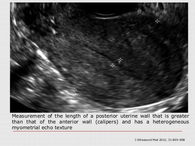
What does a heterogeneous uterus mean-–808 807 Sakhel and Abuhamad—Sonography of Adenomyosis Figure 5 Measurement of the length of a posterior uterine wall that is greater than that of the anterior wall (calipers) and has a heterogeneous myometrial echo texture Figure 430/8/ The endometrium is the lining of the uterus The endometrium is well seen on ultrasound exams and it's thickness is usually measured It is seen a stripe that is brighter than the surrounding uterine tissue it usually appears smooth and of similar consistency throughout In patients who are postmenopausal the thickness cutoff for abnormal is


Intrauterine Bony Fragments An Unexpected Finding In The Hysterectomy Specimen
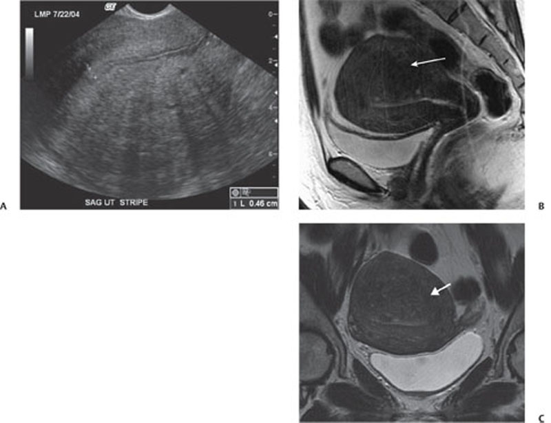
Transabdominal ultrasound using a 51MHz phased array probe in the transverse plane 2cm above pubic symphysis A large hypoechoic structure within the uterus is seen displacing the bladder superoanteriorly with posterior acoustic enhancementGeneral Use 5 MHz endocavitary probe (high frequency, low penetration) Apply surgical lubricant inside and outside probe cover Place patient in lithotomy position Gently advance probe into vaginal canal and position adjacent to cervix May be more comfortable for patient to insert probe into vagina herself Heterogeneous myometrium is more prevalent and consistent indicator for the diagnosis of adenomyosis by ultrasound Though enlarged uterus is almost invariably present in cases of adenomyosis (93%), yet it is totally nonspecific and is also present in multiparous females (see Figure 3, Figure 4) Download Download fullsize image Figure 3
Overview Leiomyomas of the uterus (or uterine fibroids) are benign tumors that arise from the overgrowth of smooth muscle and connective tissue in the uterus Histologically, a monoclonal proliferation of smooth muscle cells occursIt may mean that a fibroid or cyst was found during the ultrasound Nearly all of these are small and cause no symptoms or problems, but you need to discuss this finding with your doctor What is a heterogeneous uterus?1/3/16 On ultrasonography, most uterine leiomyomas typically appear as welldefined, solid masses Their echogenicity is usually similar to that of the myometrium, but sometimes they are hypoechoic They often show some posterior acoustic shadowing Variants of leiomyomas occur when they undergo cystic degeneration, hyalinization, or calcification
An area of irregularity in the uterus that is not a fibroid;Changes in blood flow patterns in the wall of the6/6/03 Ultrasound cannot reliably rule out retained placental tissue and especially a moderate endometrial thickness of 2–5 mm is not diagnostic 16 Consequently, to make a diagnosis of uterine AVM based only on color Doppler findings could result in overdiagnosis;


Dr Aimee Maceda On Adenomyosis Progressive Radiology


The Endometrial Echo Revisited Have We Created A Monster American Journal Of Obstetrics Gynecology
9/7/17 Ultrasound Evaluation of Myometrium The myometrium is the muscular tissue of the uterus and the cervix, which encloses the uterine cavity and its lining, the endometrium The myometrium is generally isoechoic (similar to the liver) and homogeneous The myometrial echogenicity, thickness, contour and presence of any mass or cysts are notedHace 2 días Introduction Uterine fibroids, also known as leiomyomas or myomas, are the commonest uterine neoplasms They are benign tumors of smooth muscle origin, with varying amounts of fibrous connective tissue Fibroids usually arise in the myometrium but may occasionally be found in the cervix, broad ligament or ovaries1,2 They are multiple in up toVaginal ultrasound probes may be inserted into the vagina to obtain an image of the uterus If the anteflexed uterus does cause problems, patients have several options for addressing it The degree of flexion may be mild, and it could be possible to use massage and bodywork along with muscle exercises or pessaries to push it back into a more neutral position
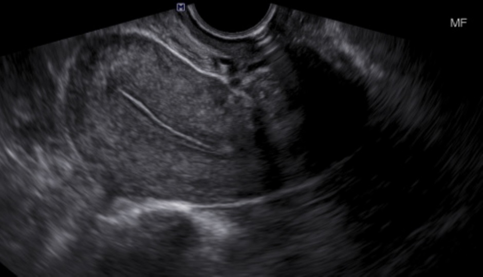


Uterine Imaging Article


Pelvic Pain Overlooked And Underdiagnosed Gynecologic Conditions Radiographics
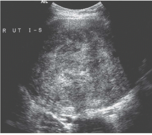
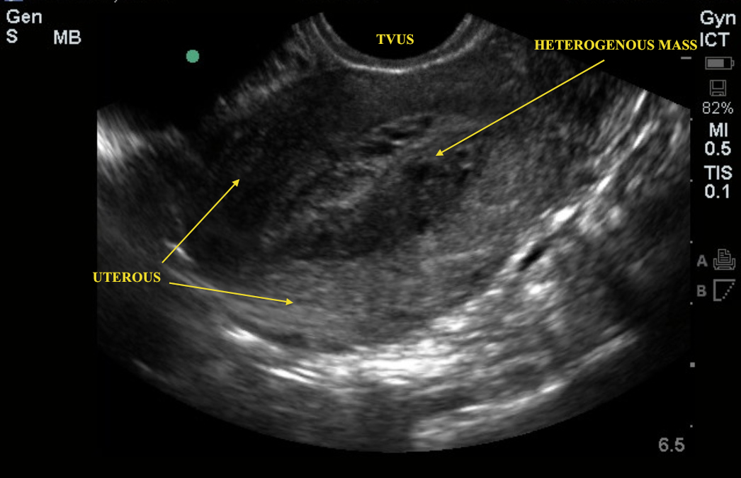
I had a ct scan and it said "the uterus is heterogeneous with low density within the endometrium and cervix the low density at the cervix measures 25 x 18 cmThe overies are unremarkable the left overy measures 27 x 27 cm and contains a 14 cm follicle the right ovary measures 36 x 15 cm and contains several follicles there is a small amount of fluid in the right adnexa a pelvic sono29/8/16 Transverse US image of the uterus in a 48yearold woman with rapid uterine enlargement shows a heterogeneous mass with prominent cystic areas expanding the uterus This proved to be a leiomyosarcoma However, cystic degeneration in a benign leiomyoma may have an identical appearanceDr Jeff Livingston answered Obstetrics and Gynecology 22 years experience
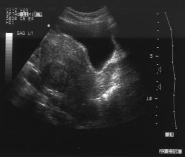


Uterine Leiomyoma Fibroid Imaging Overview Radiography Computed Tomography


Ultrasound Scans Revealed A Large Heterogeneous Mass Occupying The Download Scientific Diagram
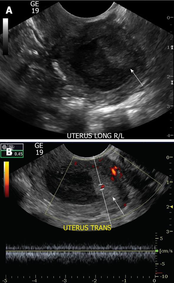
A differential thickness of the walls of the uterus;(A) Transvaginal pelvic ultrasound with color Doppler demonstrates a heterogeneous hypoechoic/isoechoic lesion arising from the myometrium (long arrow) and distorting the endometrial cavity (B) SHG demonstrates a broadbased hypoechoic lesion protruding into the endometrial canal with areas of acoustic shadowing (arrowhead) but preserving the echogenic29/3/15 Ultrasound findings that your doctor may use to help make a likely diagnosis of adenomyosis include Thickening of the area between the lining and the muscle layers in the uterus;


The Assessment Diagnosis And Causes Of Endometrial Cancer Empowered Women S Health
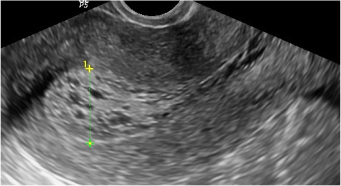


Role Of Transvaginal Ultrasound In Detection Of Endometrial Changes In Breast Cancer Patients Under Hormonal Therapy Egyptian Journal Of Radiology And Nuclear Medicine Full Text
Vaginal ultrasound showed a cystic area in my uterus Does this mean cancer No vaginal bleeding but a clear discharge on and off Belly is swollen toAnswer 'Heterogenous uterus' is a description used to describe the appearance of the uterus after an ultrasound exam is done All that this means is that the ultrasound appearance of the uterus is not totally uniform The two most common causes of heterogenous uterus are uterine fibroids, which are benign muscular growths in the uterine wall, andUterine fibroids are the most common benign uterine tumour and most common pelvic tumour in women Most are asymptomatic;


Patient Number 2 Transvaginal Ultrasound Enlarged Heterogeneous Download Scientific Diagram


A Woman With Urinary Incontinence Nejm Resident 360 Meta Property Twitter Image Content Resident360files Nejm Org Image Upload C Fit F Auto H 1 W 1 V U8buf4o8mgdxgmfcczjk Png Meta Property Og Image Content
An Ultrasound Review of PELVIC PATHOLOGY Judi M Bender MD Pathology to be covered •Uterus •Cervix •Ovary •Adnexa •Appendicitis • Enlarged heterogenic uterus • Hypoechoic/heterogenic lesion • Acoustic attenuation • Focal calcifications/ calcified rim–627 J Ultrasound Med 12;Ultrasound (US) is the accepted primary modality for the evaluation of abnormal uterine bleeding Sonohysterogram (SonoHSG) and magnetic resonance imaging (MRI) are useful for problem solving and when US is indeterminate
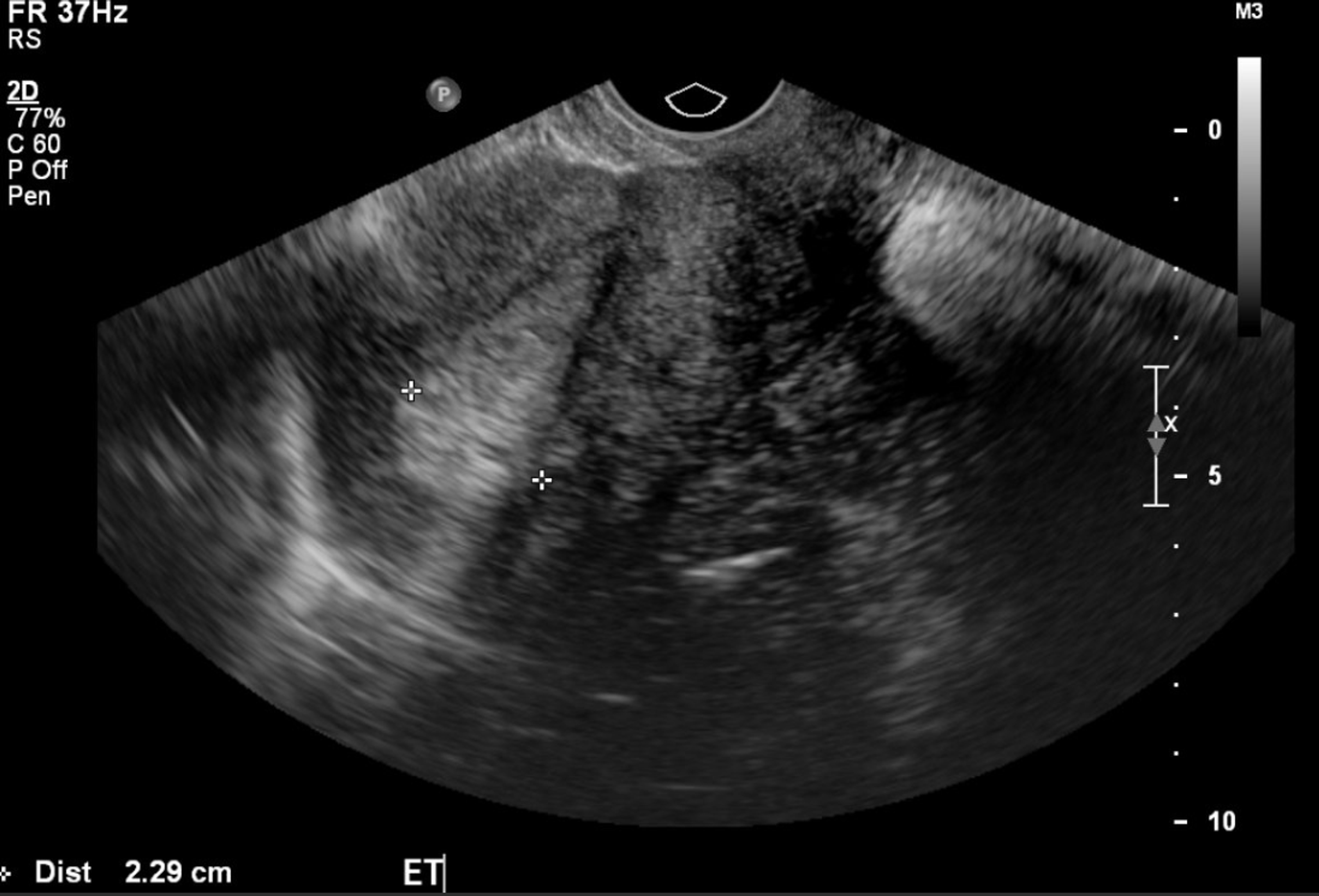


Cureus Ciliated Cell Variant Of Endometrial Carcinoma In An Adenomyoma In Uterus


Imaging The Endometrium A Pictorial Essay Sciencedirect
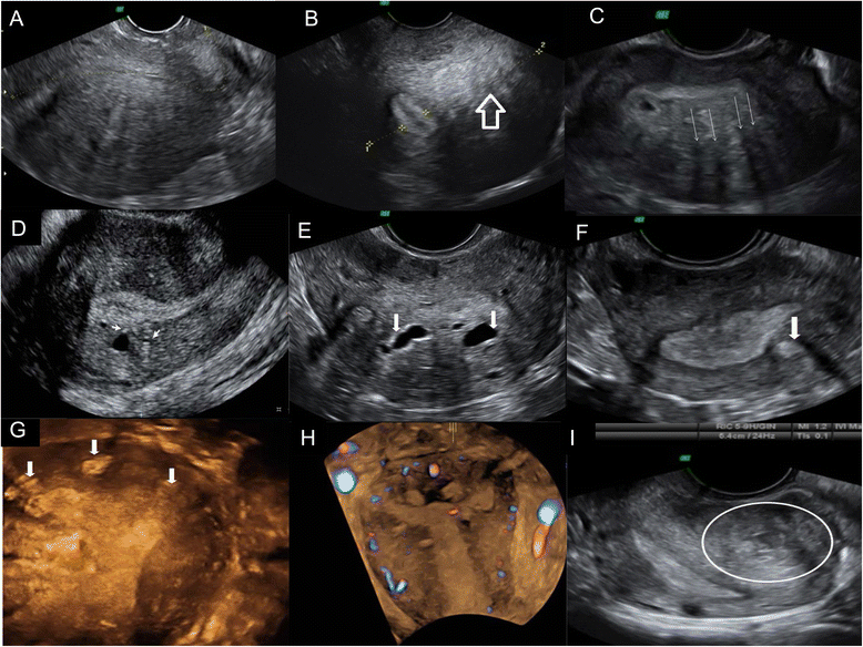
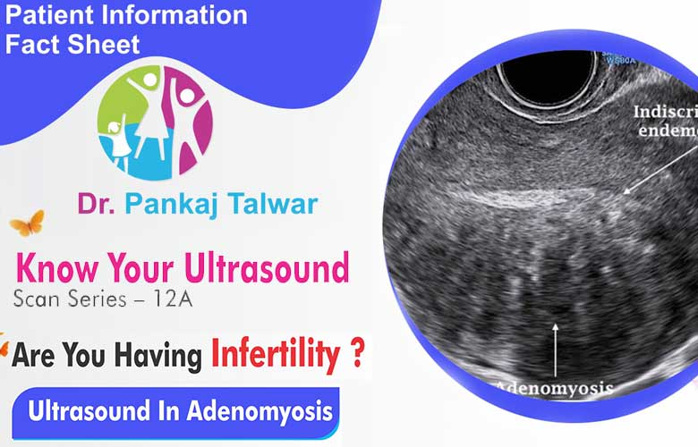
All patients with adenomyosis had a mottled heterogeneous appearing uterus, 95% had a globular uterus, % had small myometrial lucent areas, and % had an indistinct endometrial stripe Eight patients (156%) who had been suspected of having adenomyosis by pelvic sonography did not have adenomyosis reported in the pathology specimen1/5/06 Objective The purpose of this presentation is to show the imaging findings of the common and uncommon variants of adenomyosis as seen on sonography and magnetic resonance imaging (MRI) Methods A 328/3/ A heterogeneous uterus is a term used to describe the appearance of the uterus after an ultrasound is conducted It simply means that the uterus is not totally uniform in appearance during the ultrasound According to MedHelp, there are two common causes of a heterogeneous uterus uterine fibroids and adenomyosis


The Role Of Transvaginal Ultrasound Or Endometrial Biopsy In The Evaluation Of The Menopausal Endometrium American Journal Of Obstetrics Gynecology


Sonography Of Adenomyosis
However, can present with excessive uterine bleeding, symptoms secondary to pressure on bladder and rectum, and, less often, distortion of the uterine cavity, leading to miscaThe ultrasound is from a 70 year old post menopausal female who presents with an enlarged uterus The endometrial stripe is enlarged and is filled with fluid and an enhancing soft tissue mass consistent with an endometrial carcinoma Note blood flow as depicted by Doppler exam (c) characterizing the soft tissue as tumor rather than a clot I did an ultrasound and the results show uterus is heterogeneous and is 121 x 92 x cm endometrium measures 7 mm in ap thickness, left ovary 79 cc and right ovary 33 cc 3 fibroids 52 x 37 cm, 71 x 57 & 68 cm what does all of this mea?


Venetian Blind Shadowing On Ultrasound Semantic Scholar


Journals Sagepub Com Doi Pdf 10 1177
Ultrasound of the uterus with adenomyosis may reveal the organ to be of normal size or enlarged The echo texture of the affected regions is different from that of normal myometrium ( Fig 1911 );Uterus Fibroid tumors of the uterus are often found during ultrasound exams They are benign but may be hypoechoic on a sonogramThe uterus is borderline enlarged and shows heterogeneous echotexture, which is nonspecific Abnormal tissues such as cystic nodules, adenomas and cancerous tumors appear as heterogeneous structures in thyroid ultrasound imagery Cancer of the Uterus Endometrial Cancer What is adenomyosis or bulky uterus?


Academic Oup Com Humupd Article Pdf 4 4 337 Pdf


129 Adenomyosis Radiology Key
Hypoechoic myometrial striations have been described as wellEndometrial polyps are found in up to 39% of women with abnormal premenopausal bleeding and 2128% of women with postmenopausal bleeding Most endometrial polyps are benign but between 24% of them are premalignant or malignant Polyps are often suspected on a transvaginal ultrasound ButUltrasound Obstet Gynecol 03;


Assessment Of The Bovine Uterus With Endometritis Using Doppler Ultrasound Biorxiv


Diagnostics Free Full Text Differential Diagnosis Of Endometriosis By Ultrasound A Rising Challenge Html
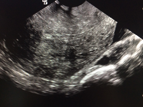
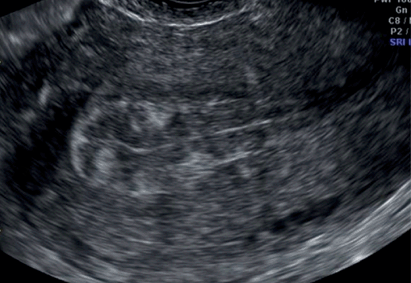
On ultrasound imaging, the presence of fibroids can usually be detected by heterogeneous enlargement of the uterus due to the presence of welldemarcated, hypoechoic masses within the myometrium However, Fibroids may also appear isoechoic or hyperechoicStatus, normal adult uterus measures approximately 7290cm long, 4560cm wide and 535 deep(1) Uterine size has been found to be parity related and not age related(2) After menopause, uterine sizes decrease due to reduction of ovarian hormone Pelvic ultrasound is12/9/18 Muscular hyperplasia and hypertrophy cause focal or diffuse myometrial thickening and globular uterine enlargement, often with thin "venetian blind" shadows The combination of these findings results in a heterogeneous myometrium, with blurring of the endometrial border


Www Dsjuog Com Doi Dsjuog Pdf 10 5005 Jp Journals 1506


Abnormally Thickened Endometrium Differential Radiology Reference Article Radiopaedia Org
The main method of diagnosing the condition of the uterus cavity is still ultrasound, which should be carried out after menstruation A heterogeneous endometrium is often detected in the research process, and such a pathological condition may develop for a number of reasonsIn a recent CT scan for kidney stones, it said I had a "heterogeneous uterus" with left and right "adnexal cysts" I have polycystic ovarian syndrome, so the cysts are not really a surprise, but I don't know what a heterogeneous uterus is It didn't mention any mass or fluid, just recommended an ultrasoundJournal of Ultrasound in Medicine Volume 25, Issue 5 p Image Presentation AdenomyosisCommon and Uncommon Manifestations on Sonography and Magnetic Resonance Imaging Sheetal Chopra MBBS, DNB, Thomas Jefferson University Hospital, Philadelphia, Pennsylvania USA


What Are The Most Reliable Signs For The Radiologic Diagnosis Of Uterine Adenomyosis An Ultrasound And Mri Prospective Sciencedirect


Adenomyosis Glowm
Why does blood pressureAdenomyosis (or uterine adenomyosis) is a common uterine condition of ectopic endometrial tissue in the myometrium, sometimes considered a spectrum of endometriosis Although most commonly asymptomatic, it may present with menorrhagia and dysmenorrhea Pelvic imaging (ie ultrasound, MRI) may show characteristic findingsHeterogeneous refers to a structure with dissimilar components or elements, appearing irregular or variegated For example, a dermoid cyst has heterogeneous attenuation on CT It is the antonym for homogeneous , meaning a structure with similar components


Pelvic Ultrasound Demonstrating A Thickened Heterogeneous Endometrial Download Scientific Diagram


Adenomyosis A Sonographic Diagnosis Radiographics
Hence the conclusion in some recent reports 2 , 3 , 12 that uterine AVMs are much more common than previously thoughtA heterogeneous myometrial echotexture on ultrasound is usually a nonspecific finding, although it has been described with uterine adenomyosis What is a heterogeneous appearance?Myometrium is heterogeneous features Any disease can be prevented in time turning to the doctor Any disease can be overcome with proper diagnosis and timely treatment Today, women of reproductive age are increasingly faced with the problem of infertility due to various diseases, hormonal disorders or frequent abortions


The Accuracy Of Transvaginal Ultrasound And Uterine Artery Doppler In The Prediction Of Adenomyosis Sciencedirect


References In Imaging For Uterine Myomas And Adenomyosis Journal Of Minimally Invasive Gynecology
29/5/ Myometrium is the muscular wall of the uterus or it is also called as the middle layer of the uterine wall The normal condition of a uterus is called as homogeneous myometium In other words we can say that, a homogeneous myometrium is free of Lumps, voids which is the normal condition of the uterus4/3/15 Heterogeneous myometrial echotexture on ultrasound is a nonspecific finding but can be associated with adenomyosis of the uterus or fibroids If you were told that endometrial cells were found on the surface of the uterus, it mostly meansThe best triple combined variables were 'bulky uterus', 'heterogeneous myometrium' 'ill definition of the endometrialmyometrial interface' (sensitivity 38%, specificity 93%) Conclusion Transvaginal ultrasound is highly specific for diagnosing uterine adenomyosis, providing a costeffective and readily available alternative to MRI


Imaging The Endometrium A Pictorial Essay Sciencedirect


When Would You Suspect Fibroid Uterus
Myometrium is the muscular layer of the uterus which allows the uterus to expand in size during pregnancy Echo texture refers to the appearance of the myometrium on ultrasound examination So a normal myometrial echotexture is a good thing This6/3/18 The normal postmenopausal endometrium will appear thin, homogenous and echogenic Endometrial cancer causes the endometrium to thicken, appear heterogeneous, have irregular or poorly defined margins, and show increased color Doppler signals 3D Ultrasound Normal Endometrial Thickness26/3/ Inhomogeneous uterus texture refers to the appearance of focal masses giving a nonuniform heterogeneous surface of the uterus exterior wall, according to National Center for Biotechnology Information The two most common causes of inhomogeneous uterus are uterine fibroids and adenomyosis Uterine fibroids are benign tumors of muscular or


Adenomyosis Chapter 16 Ultrasonography In Reproductive Medicine And Infertility


Diseases Of The Uterus Radiology Key
1/5/03 Any significant deviation from a woman's established menstrual pattern may be considered abnormal uterine bleeding, and several factors direct evaluation of a patient with such bleeding Premenopausal disorders that are well evaluated with ultrasound (US) include endometriosis, adenomyosis, and leiomyomasIt may be heterogeneous or more echogenic than myometrium;


Www Aium Org Misc Soundjudgment6 Pdf


Abnormally Thickened Endometrium Differential Radiology Reference Article Radiopaedia Org


Choriocarcinoma A Rapidly Progressive Unusual Tumor Eurorad


The Normal Uterus


Academic Oup Com Humupd Article Pdf 4 4 337 Pdf


Imaging The Endometrium A Pictorial Essay Sciencedirect


Imaging The Endometrium Disease And Normal Variants Radiographics
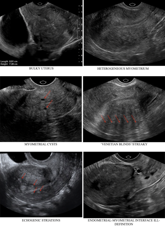


Accuracy Of Findings In The Diagnosis Of Uterine Adenomyosis On Ultrasound Springerlink


Intrauterine Bony Fragments An Unexpected Finding In The Hysterectomy Specimen


Adenomyosis Common And Uncommon Manifestations On Sonography And Magnetic Resonance Imaging Chopra 06 Journal Of Ultrasound In Medicine Wiley Online Library


View Image


Tamoxifen Associated Changes Endometrial Hyperplasia Can Occur In


Q Tbn And9gcqgiazvdncyeycr Vpzzfvtzwzja8iisrxbr2jscdcm8pq Eitf Usqp Cau


Www Dsjuog Com Doi Dsjuog Pdf 10 5005 Jp Journals 1594


What Are The Most Reliable Signs For The Radiologic Diagnosis Of Uterine Adenomyosis An Ultrasound And Mri Prospective Sciencedirect


Ultrasound Evaluation Of Myometrium Obgyn Key
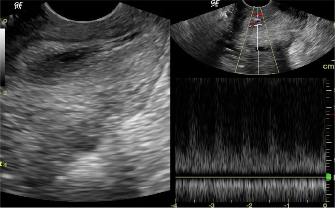


Role Of Transvaginal Ultrasound In Detection Of Endometrial Changes In Breast Cancer Patients Under Hormonal Therapy Egyptian Journal Of Radiology And Nuclear Medicine Full Text


Early Diagnosis Of Spontaneous Heterotopic Pregnancy Successfully Treated With Laparoscopic Surgery Bmj Case Reports


3


Endometrial Thickness Predicts Intrauterine Pregnancy In Patients With Pregnancy Of Unknown Location Moschos 08 Ultrasound In Obstetrics Amp Gynecology Wiley Online Library


Ultrasound Characteristics Of Endometrial Cancer As Defined By International Endometrial Tumor Analysis Ieta Consensus Nomenclature Prospective Multicenter Study Epstein 18 Ultrasound In Obstetrics Amp Gynecology Wiley Online Library


Radiological Category Genitourinary Principal Modality 1 Ultrasound Principal
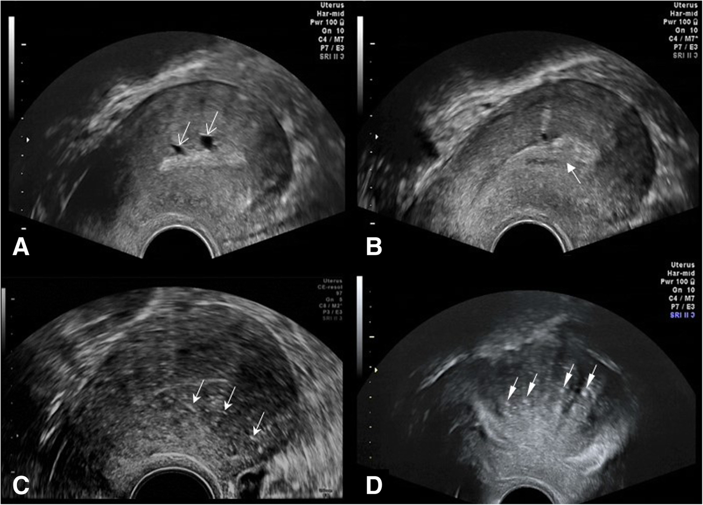


Management Of Uterine Adenomyosis Current Trends And Uterine Artery Embolization As A Potential Alternative To Hysterectomy Insights Into Imaging Full Text


How To Diagnose Adenomyosis Why Are We Missing It Sydney Fibroid Clinic


Www Bmus Org Static Uploads Resources Uterus Endometrium Common Uncommon Pathologies Bmus York Pdf


Figure 1 A Case Of Adenosarcoma Of The Uterus


Role Of Transvaginal Sonography And Magnetic Resonance Imaging In The Diagnosis Of Uterine Adenomyosis Fertility And Sterility


Imaging The Endometrium Disease And Normal Variants Radiographics


Ultrasound Evaluation Of Myometrium Obgyn Key


Transvaginal Ultrasound Of A Normal Uterus 5 Days Postpartum Transverse Image Shows Echogenic Foci In The Endometri Transvaginal Ultrasound Uterus Image Shows


Adenomyosis Adenomyosis And Pregnancy Ivf1


Www Glowm Com Pdf Ultrasound In Obstetrics And Gynecology Chapter11 Pdf


Gynecologic Ultrasound Radiologic Clinics


Preimplantation 3d Ultrasound Current Uses And Challenges


Association Of 2d And 3d Transvaginal Ultrasound Findings With Adenomyosis In Symptomatic Women Of Reproductive Age A Prospective Study Clinics
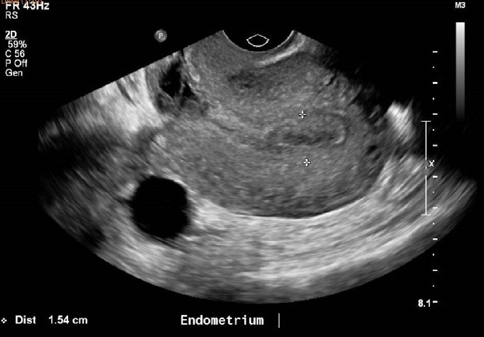


Endometrial Hyperplasia As A Cause Of Secondary Postpartum Hemorrhage In A Post Caesarean Section Patient Goh Journal Of Medical Cases


The Assessment Diagnosis And Causes Of Endometrial Cancer Empowered Women S Health
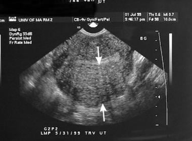


Adenomyosis Imaging Practice Essentials Magnetic Resonance Imaging Ultrasonography


When Would You Suspect Fibroid Uterus


Doppler Ultrasound In Gynaecology Chapter 16 Gynaecological Ultrasound Scanning


Q Tbn And9gcq4n9kjisge1c2im3e Dttnlw8eipza2kkfxe6rmcfb2il5dfe6 Usqp Cau
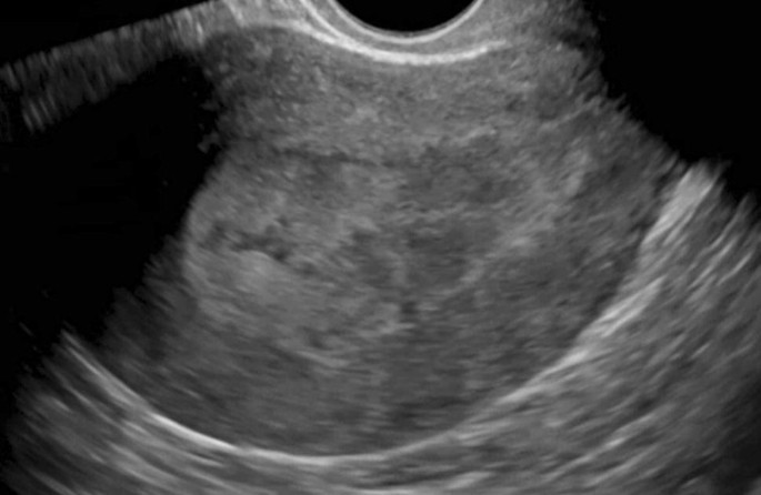


Recent Updates In Female Pelvic Ultrasound Springerlink


Jaypeedigital Ebook Reader


Adenomyosis


Www Bmus Org Static Uploads Resources Uterus Endometrium Common Uncommon Pathologies Bmus York Pdf


Endometrial Hyperplasia Radiology Reference Article Radiopaedia Org


Retained Products Of Conception Radiologypics Com
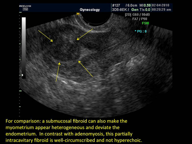


Uterine Adenomyosis Noninvasive Diagnosis Mdedge Obgyn
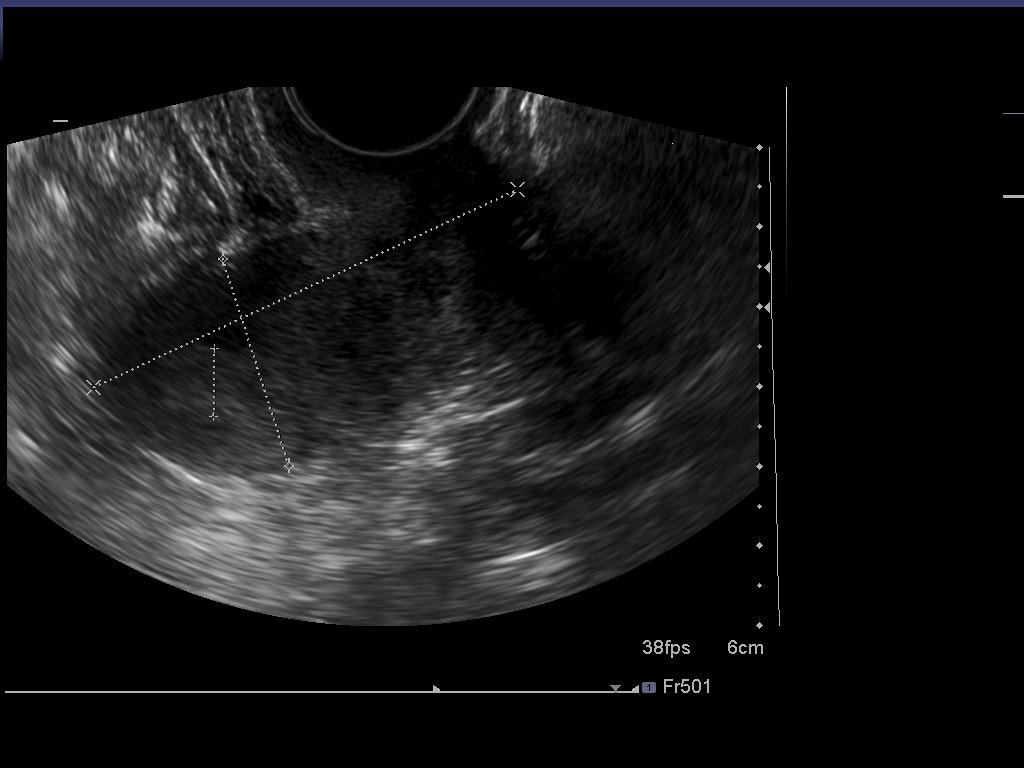


Epos Trade


1


Adenomyosis Radiology Case Radiopaedia Org


Pelvic Pain Overlooked And Underdiagnosed Gynecologic Conditions Radiographics


Retained Products Of Conception An Atypical Presentation Diagnosed Immediately With Bedside Emergency Ultrasound


Adenomyosis Chapter 16 Ultrasonography In Reproductive Medicine And Infertility


Sonohysterography Principles Technique And Role In Diagnosis Of Endometrial Pathology


Adenomyosis In Infertile Women Prevalence And The Role Of 3d Ultrasound As A Marker Of Severity Of The Disease Reproductive Biology And Endocrinology Full Text


Retained Products Of Conception An Atypical Presentation Diagnosed Immediately With Bedside Emergency Ultrasound


When Would You Suspect Fibroid Uterus


Adenomyosis Common And Uncommon Manifestations On Sonography And Magnetic Resonance Imaging Chopra 06 Journal Of Ultrasound In Medicine Wiley Online Library


Www Aium Org Misc Soundjudgment6 Pdf


Passing Clots Emory School Of Medicine


Abdominal Ultrasound With Heterogeneous Contents In The Uterine Cavity Download Scientific Diagram


Abdominal Pain In Nonpregnant Female Patients 14 04 06 Ahc Media Continuing Medical Education Publishing


Ultrasound In Adenomyosis Fertility Treatment Center Delhi Ncr


Adenomyosis Collection Of Ultrasound Images


Www Bmus Org Static Uploads Resources Uterus Endometrium Common Uncommon Pathologies Bmus York Pdf


Pelvic Pain Overlooked And Underdiagnosed Gynecologic Conditions Radiographics


42 Year Old Woman With Abnormal Uterine Bleeding Mdedge Obgyn


Sonographic Assessment Of Endometrial Pathology Chapter 6 Gynaecological Ultrasound Scanning


Www Bmus Org Static Uploads Resources Uterus Endometrium Common Uncommon Pathologies Bmus York Pdf


Adenomyosis Radiology Reference Article Radiopaedia Org


Sagittal View Of The Uterus On Trans Vaginal Ultrasound A Bulky Download Scientific Diagram
コメント
コメントを投稿